Dermatological ServicesDermatoscopy: Non-Invasive Skin Examination and Diagnosis

Dermatoscopy: Non-Invasive Skin Examination and Diagnosis in Latvia

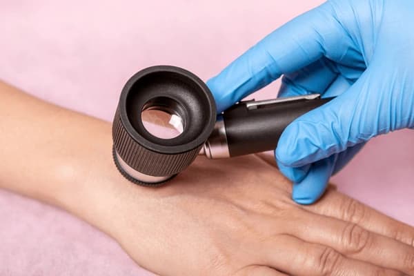
Gloved doctor's hand examining the skin on a patient's hand with a dermatoscope during a mole check.

Need an online dermatologist for dermatoscopy in Latvia? iDerma's doctors review your photos and reply within 24 hours — from 45 €.

Dermatoscopy – A Method for Examining the Surface of the Skin

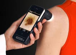
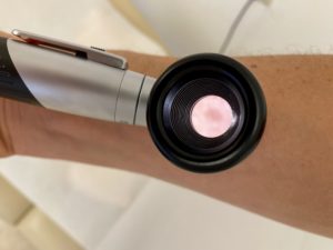
Dermatoscopy is a surface-level skin examination method using a special device called a dermatoscope, which allows for microscopic evaluation of the skin. This method is both informative and easy to apply in daily clinical practice.

Most commonly, various skin lesions are examined under the dermatoscope. The following aspects of a lesion are evaluated:

  • structure (symmetry, uniformity)

  • color

  • pigmentation

  • the arrangement of blood vessels

  • border characteristics (smoothness, color variation)

  • other changes such as ulceration


Suspicious features for skin cancer may include:

  • irregular structure

  • uneven color

  • uneven pigmentation, visible changes in pigment tone

  • dense or irregularly shaped blood vessel networks

  • irregular, multi-colored borders

  • visible ulceration or bleeding in the lesion


Using dermatoscopy, it is possible to detect or suspect:

  • melanoma

  • basal cell carcinoma

  • various types of moles

  • seborrheic keratosis

  • hemangioma


ARTICLE_GIF


Other uses of dermatoscopy:

  • to determine the type of hair loss (trichoscopy) by examining hair and surrounding scalp

  • to assess non-pigmented lesions

  • to diagnose scabies

  • for differential diagnosis between chronic inflammatory skin diseases, such as:

    • psoriasis

    • eczema

    • lichen planus

  • to examine capillaries in the nail bed

  • to locate small embedded foreign bodies, such as splinters

Still unsure?

A dermatologist writes a plan made for your skin.

Not another off-the-shelf cream — a certified specialist’s diagnosis and personal treatment plan, within 24 hours.

Start your consultation
Personal treatment plan
24h
Diagnosis
Treatment plan
Prescriptions
iDerma
Board-certified dermatologist

Article by

Anna Tunkeviča

Medical content reviewed by

Andra Šukienė(Dermatologist)

Our other articles

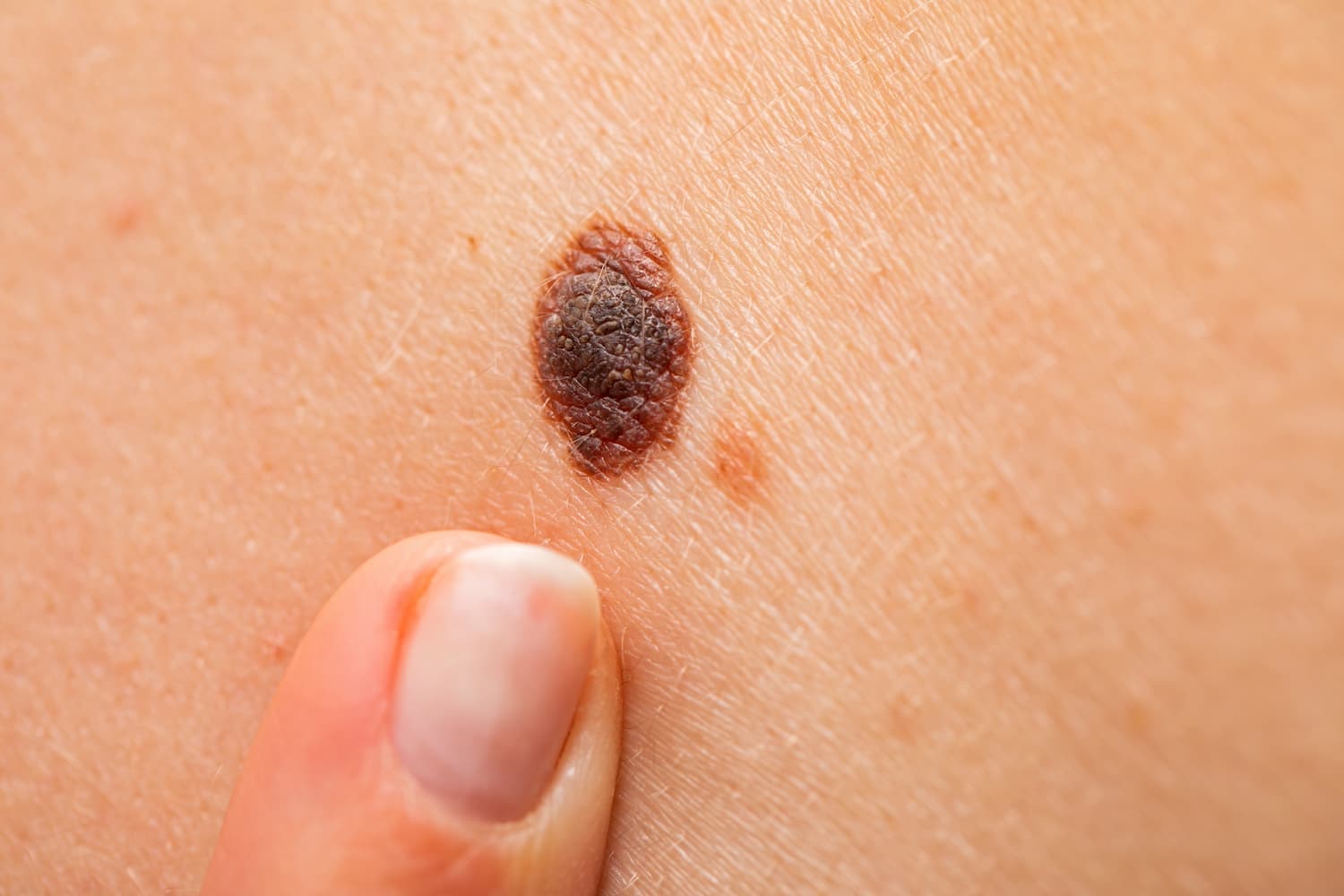
Raised dark brown melanocytic nevus on the skin, the kind a dermatologist assesses for benign features.

Skin moles: how to tell if they're benign or dangerous

Benign melanocytic nevi are harmless moles, but changes in size, shape, or color may signal risk. Learn when to monitor or remove them.
Gloved specialist using a cryotherapy device to treat a skin lesion on a patient's hand.

Cryotherapy: minimally invasive skin lesion removal

Cryotherapy is a safe and fast method for removing skin lesions using cold agents like liquid nitrogen. Effective for benign growths with minimal downtime.
Large asymmetric brown-black skin lesion with irregular borders and uneven colour, suspicious for melanoma

Melanoma – skin cancer symptoms, causes & treatment options

Melanoma is the deadliest form of skin cancer. Discover how to spot early signs, when to see a doctor, and what treatments are available.
Pearly pink nodule on the skin beside the eye, typical of basal cell carcinoma, a common form of skin cancer.

Basal cell carcinoma: causes, risk factors, symptoms, and treatment

Basal cell carcinoma is the most common skin cancer. It rarely spreads but requires treatment to prevent recurrence and cosmetic complications.